


Nanofluidic channel-resonant cavity structure for measuring micro-displacement of fluorescent substances
-
摘要: 本文提出了一种纳流通道-谐振腔耦合结构,用于实现对荧光物质微位移的检测。在本文中,首先,使用时域有限差分法,研究了量子点偏振态及结构参数对荧光与结构耦合效果的影响,进而对结构进行优化;然后,通过测量耦合结构输出光功率的变化,实现对荧光物质微位移的检测;最后,对影响传感灵敏度的因素进行研究。结果表明,相比传统方法,纳流通道-谐振腔耦合结构的折射率处于2.8~3.3之内时,该结构都可以实现对荧光物质微位移的高精度准确传感,并且通过减小纳流通道与谐振腔的间距可进一步提高传感灵敏度。Abstract: In order to measure the micro-displacement of a fluorescent substance, we propose a nanofluidic channel-resonant cavity structure. Firstly, by using the Finite-Difference Time-Domain (FDTD) method, the influences of the quantum dot’s polarization state and structural parameters on the coupling effect of fluorescence and structure are studied and the structure is optimized. Then, the micro-displacement of the fluorescent substance is detected by measuring the change in the optical power output of the coupled structure. Finally, the factors affecting the sensitivity of the sensors are studied. The results show that, compared with the traditional method, when the refractive index of the nanofluidic channel-resonant cavity coupling structure is in the 2.8~3.3 range, the structure can sense of the micro-displacement of a fluorescent substance with high accuracy. The results also show that the sensing sensitivity can be further improved by reducing the distance between the nanofluidic channel and the resonant cavity.
-
Key words:
- nanofluidic channel /
- resonant cavity /
- quantum dots /
- micro-displacement
-
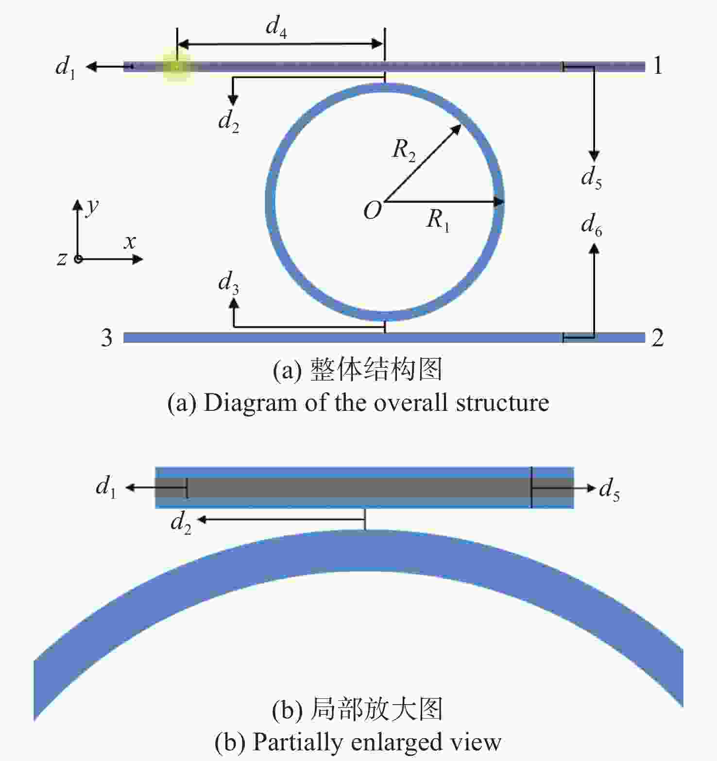
图 1 纳流通道-谐振腔耦合结构二维模型图
Figure 1. Two-dimensional model diagram of a nanofluidic channel-resonant cavity structure
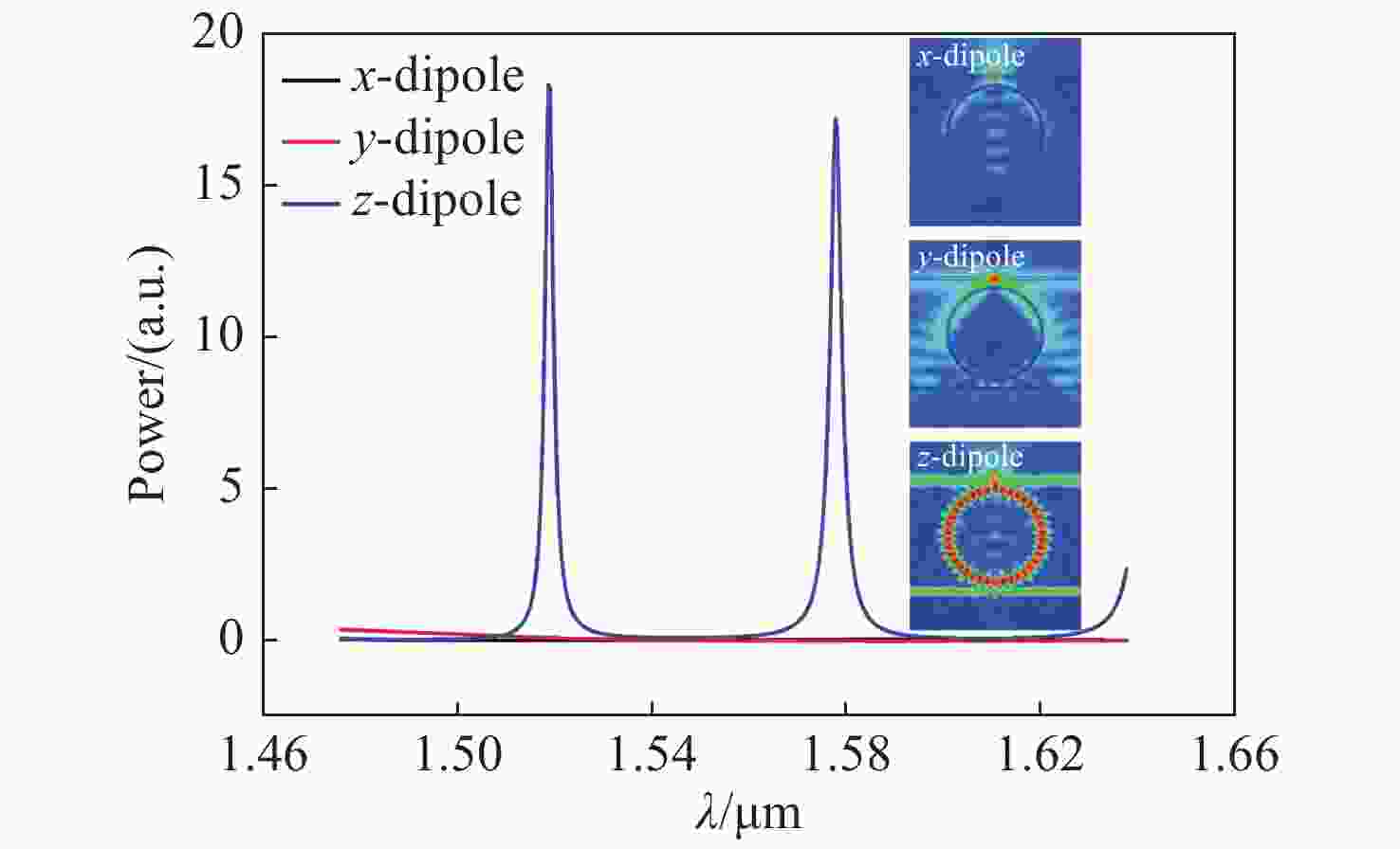
图 3 偶极子光源偏振方向不同时的耦合效果曲线和电场分布图
Figure 3. Coupling effect curves and electric field distributions of dipole source with different polarization directions

图 4 纳流通道及下波导与谐振腔间距不同时的耦合效果曲线
Figure 4. Coupling effect curves when the distance between the microfluidic channel, the lower waveguide and the resonant cavity are different
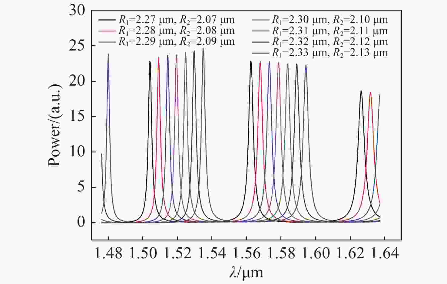
图 5 不同谐振腔大小时的耦合效果曲线
Figure 5. Coupling effect curves when the cavity size is different

图 6 纳流通道参数不同时的耦合效果曲线
Figure 6. Coupling effect curves when the microfluidic channel parameters are different
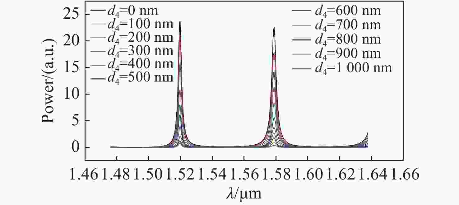
图 7 不同量子点位置时端口2的光功率曲线
Figure 7. Optical power curves at port 2 when the quantum dot position changes

图 8 量子点处于不同位置时的电场分布
Figure 8. Electric field distributions when quantum dots are in different positions

图 9 量子点与结构中心水平距离d4变化时端口2的峰值功率曲线
Figure 9. Peak power curve of port 2 when the horizontal distance d4 between the quantum dot and the center of the structure changes
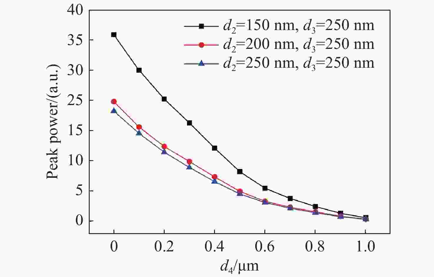
图 10 结构间距不同,量子点与结构中心水平距离d4变化时端口2的峰值功率曲线
Figure 10. Peak power curves of port 2 varying with d4, the horizontal distance between the quantum dot and the center of the structure, at different structure spacing
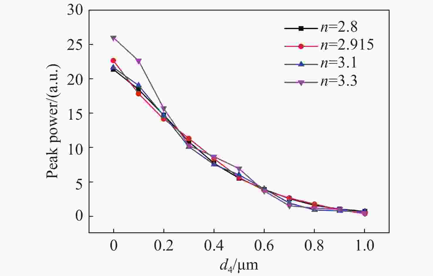
图 11 不同结构折射率时,量子点与结构中心水平距离d4变化时端口2的峰值功率曲线
Figure 11. Peak power curves of port 2 varying with d4, the horizontal distance between the quantum dot and the center of the structure, at different refractive indexs
-
[1] 陈飘飘, 邢怡晨, 刘洋, 等. 基于DNA QDs@PDA荧光共振能量转移的半胱氨酸传感器[J]. 分析化学,2020,48(1):83-89.CHEN P P, XING Y CH, LIU Y, et al. DNA Quantum Dots@Polydopamine as a fluorescent sensor for cysteine detection based on fluorescence resonance energy transfer effect[J]. Chinese Journal of Analytical Chemistry, 2020, 48(1): 83-89. (in Chinese) [2] MEDINTZ I L, UYEDA H T, GOLDMAN E R, et al. Quantum dot bioconjugates for imaging, labelling and sensing[J]. Nature Materials, 2005, 4(6): 435-446. doi: 10.1038/nmat1390 [3] 杜方凯, 张慧, 谭学才, 等. 基于氮掺杂石墨烯量子点/硫化镉纳米晶电化学发光传感器检测硫化氢[J]. 分析化学,2020,48(2):240-247.DU F K, ZHANG H, TAN X C, et al. Detection of hydrogen sulfide based on nitrogen-doped graphene quantum dots/cadmium sulfide nanocrystals electrochemiluminescence sensor[J]. Chinese Journal of Analytical Chemistry, 2020, 48(2): 240-247. (in Chinese) [4] 康倩文, 张国, 柴瑞涛, 等. 基于碳纳米点荧光增强检测铝离子[J]. 分析化学,2019,47(12):1901-1908.KANG Q W, ZHANG G, CHAI R T, et al. Synthesis of carbon nanodots for detection of aluminum ion with fluorescence enhancement[J]. Chinese Journal of Analytical Chemistry, 2019, 47(12): 1901-1908. (in Chinese) [5] 陈蜜, 岳仁叶, 李智, 等. 串联的纳米传感器用于癌细胞中miRNA的超灵敏检测[J]. 分析化学,2020,48(1):40-48.CHEN M, YUE R Y, LI ZH, et al. Cascaded nanosensors for ultrasensitive detection of miRNA in cancer cells[J]. Chinese Journal of Analytical Chemistry, 2020, 48(1): 40-48. (in Chinese) [6] GUASTO J S, BREUER K S. High-speed quantum dot tracking and velocimetry using evanescent wave illumination[J]. Experiments in Fluids, 2009, 47(6): 1059. doi: 10.1007/s00348-009-0700-z [7] CUI L, ZHANG T, MORGAN H. Optical particle detection integrated in a dielectrophoretic lab-on-a-chip[J]. Journal of Micromechanics and Microengineering, 2002, 12(1): 7-12. doi: 10.1088/0960-1317/12/1/302 [8] HISHIDA K, SAKAKIBARA J. Combined planar laser-induced fluorescence–particle image velocimetry technique for velocity and temperature fields[J]. Experiments in Fluids, 2000, 29(1): S129-S140. [9] STRUBEL V, SIMOENS S, VERGNE P, et al. Fluorescence tracking and μ-PIV of individual particles and lubricant flow in and around lubricated point contacts[J]. Tribology Letters, 2017, 65(3): 75. doi: 10.1007/s11249-017-0859-z [10] VARELA S, BALAGUÉ I, SANCHO I, et al. Functionalised alginate flow seeding microparticles for use in Particle Image Velocimetry (PIV)[J]. Journal of Microencapsulation, 2016, 33(2): 153-161. doi: 10.3109/02652048.2016.1142016 [11] MEINHART C D, WERELEY S T, SANTIAGO J G. PIV measurements of a microchannel flow[J]. Experiments in Fluids, 1999, 27(5): 414-419. doi: 10.1007/s003480050366 [12] SANTIAGO J G, WERELEY S T, MEINHART C D, et al. A particle image velocimetry system for microfluidics[J]. Experiments in Fluids, 1998, 25(4): 316-319. doi: 10.1007/s003480050235 [13] JIN S, HUANG P, PARK J, et al. Near-surface velocimetry using evanescent wave illumination[J]. Experiments in Fluids, 2004, 37(6): 825-833. doi: 10.1007/s00348-004-0870-7 [14] SADR R, YODA M, ZHENG Z, et al. An experimental study of electro-osmotic flow in rectangular microchannels[J]. Journal of Fluid Mechanics, 2004, 506: 357-367. doi: 10.1017/S0022112004008626 [15] ZETTNER C, YODA M. Particle velocity field measurements in a near-wall flow using evanescent wave illumination[J]. Experiments in Fluids, 2003, 34(1): 115-121. doi: 10.1007/s00348-002-0541-5 [16] POUYA S, KOOCHESFAHANI M, SNEE P, et al. Single Quantum Dot (QD) imaging of fluid flow near surfaces[J]. Experiments in Fluids, 2005, 39(4): 784-786. doi: 10.1007/s00348-005-0004-x [17] OKAMOTO K, NISHIO S, SAGA T, et al. Standard images for particle-image velocimetry[J]. Measurement Science and Technology, 2000, 11(6): 685-691. doi: 10.1088/0957-0233/11/6/311 [18] FOREMAN M R, SWAIM J D, VOLLMER F. Whispering gallery mode sensors[J]. Advances in Optics and Photonics, 2015, 7(2): 168-240. doi: 10.1364/AOP.7.000168 [19] BUTT M A, KHONINA S N, KAZANSKIY N L. Hybrid plasmonic waveguide-assisted Metal–Insulator–Metal ring resonator for refractive index sensing[J]. Journal of Modern Optics, 2018, 65(9): 1135-1140. doi: 10.1080/09500340.2018.1427290 [20] WHITE I M, ZHU H Y, SUTER J D, et al. Refractometric sensors for lab-on-a-chip based on optical ring resonators[J]. IEEE Sensors Journal, 2007, 7(1): 28-35. doi: 10.1109/JSEN.2006.887927 [21] KWON M S, STEIER W H. Microring-resonator-based sensor measuring both the concentration and temperature of a solution[J]. Optics Express, 2008, 16(13): 9372-9377. doi: 10.1364/OE.16.009372 [22] LIU ZH H, LIU L, ZHU Z D, et al. Whispering gallery mode temperature sensor of liquid microresonastor[J]. Optics Letters, 2016, 41(20): 4649-4652. doi: 10.1364/OL.41.004649 [23] XU H T, HAFEZI M, FAN J, et al. Ultra-sensitive chip-based photonic temperature sensor using ring resonator structures[J]. Optics Express, 2014, 22(3): 3098-3104. doi: 10.1364/OE.22.003098 [24] KOCH B, YI Y, ZHANG J Y, et al. Reflection-mode sensing using optical microresonators[J]. Applied Physics Letters, 2009, 95(20): 201111. doi: 10.1063/1.3263143 [25] LI B B, CLEMENTS W R, YU X C, et al. Single nanoparticle detection using split-mode microcavity Raman lasers[J]. Proceedings of the National Academy of Sciences of the United States of America, 2014, 111(41): 14657-14662. doi: 10.1073/pnas.1408453111 [26] ZHI Y Y, YU X CH, GONG Q H, et al. Single nanoparticle detection using optical microcavities[J]. Advanced Materials, 2017, 29(12): 1604920. doi: 10.1002/adma.201604920 [27] FERN R E, ONTON A. Refractive index of AlAs[J]. Journal of Applied Physics, 1971, 42(9): 3499-3500. doi: 10.1063/1.1660760 [28] YEE K. Numerical solution of initial boundary value problems involving maxwell's equations in isotropic media[J]. IEEE Transactions on Antennas and Propagation, 1966, 14(3): 302-307. doi: 10.1109/TAP.1966.1138693 [29] CHEN ZH H, WANG Y, YANG Y B, et al. Enhanced normal-direction excitation and emission of dual-emitting quantum dots on a cascaded photonic crystal surface[J]. Nanoscale, 2014, 6(24): 14708-14715. doi: 10.1039/C4NR03851G -


 下载:
下载:


计量
- 文章访问数: 1369
- HTML全文浏览量: 323
- PDF下载量: 94
- 被引次数: 0
