


Ultrasound image segmentation based on a multi-parameter Gabor filter and multiscale local level set method
-
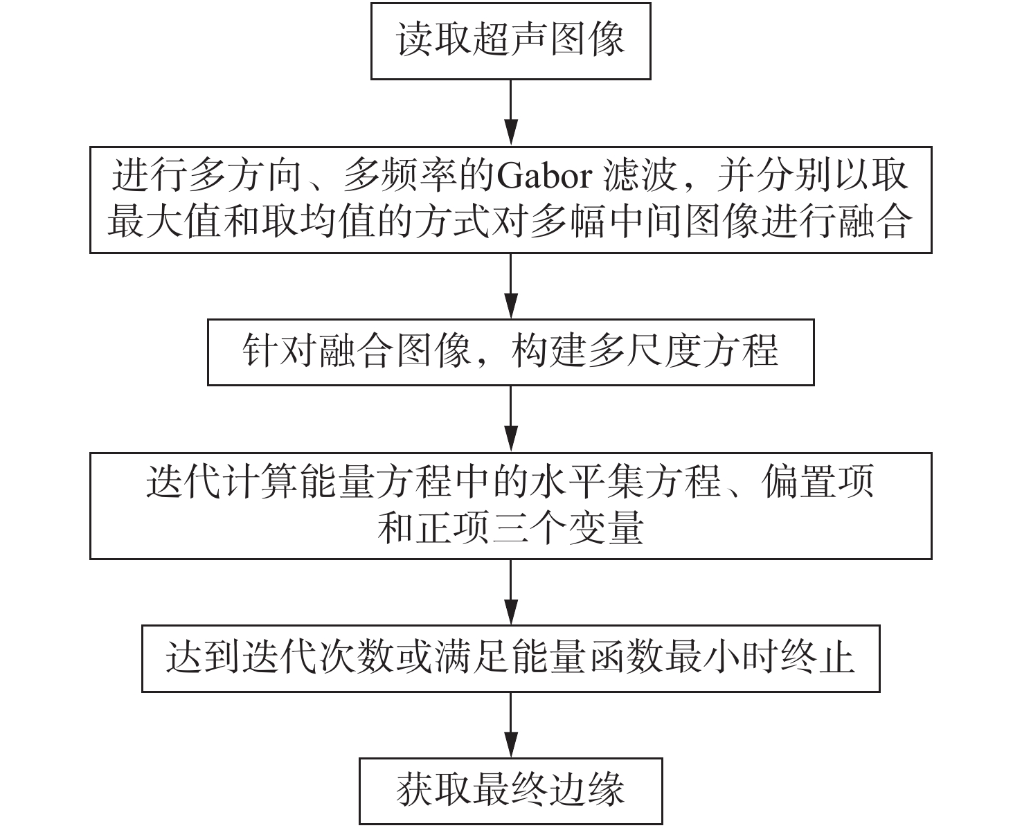
摘要: 针对超声图像边缘较弱且不连续、图像灰度分布不均的特点,提出一种基于多方向、多频率的Gabor滤波融合多尺度水平集的边缘提取算法。将超声图像成像的不连续性看作随机方向的纹理,利用Gabor滤波的方向性进行不同角度的滤波,通过最大值融合多图像,得到待分割区域和背景之间的差异且最大程度地保留原图像信息的中间图像。同时,使用多中心频率的Gabor滤波核以满足超声图像复杂的频率分布特性,并通过均值融合的方式减弱噪声的影响。再针对融合图像边缘较弱且灰度变化不均的缺陷,改进传统的局部聚类水平集方法,采用不同方差大小的高斯卷积核来适应图像不同部分的灰度变化情况,通过均值融合构造多尺度能量函数。通过在增强图像上迭代改进后的多尺度水平集函数,获取最终边缘。为验证算法的有效性,对胃部超声图像进行测试,分割结果的相关性系数和敏感性系数分别达到了0.856和0.910,相比传统局部强度聚类水平集方法分别提升了20.7%和5%。实验结果表明,该算法可以显著提高超声图像边缘提取的连续性和准确性,有效降低因超声图像灰度不均和边缘较弱造成的过分割现象。Abstract: To address the weakness and discontinuity of the edges and the uneven distribution of gray in ultrasonic images, an improved edge extraction algorithm based on a multi-parameter Gabor filter and multiscale local level set method is proposed. With the grayscale inhomogeneity of ultrasound images being regarded as texture in different directions, the directionalities of the Gabor wavelet are adopted to filter at different angles. An intermediate image is obtained to isolate the difference between each region and the background, which will allow the retention of the original image by maximizing it with a fusion method. The Gabor filter kernel with multi-center frequency meets the complex frequency distribution characteristics of ultrasound images, and the mean fusion method is used to maximize the information in the image while reducing noise influence. For the edge of the ultrasound image is weak and the grayscale is uneven, the local intensity clustering level set method is improved. A Gaussian convolution kernel template is applied with different variance sizes to fit the grayscale changes in different parts of the image. Testing the ultrasound images of a stomach show that correlation coefficient and sensitivity coefficient reaches 0.856 and 0.910, respectively, which is a 20.7% and 5% improvement over the traditional LIC algorithm, respectively. This method can satisfy the system requirements where non-contact, online, real-time, higher precision and rapid speed strong anti-jamming and stabilization are needed.
-
Key words:
- ultrasound image segmentation /
- edge extraction /
- Gabor filter /
- level set method
-

图 2 超声图像分割结果图。 (a)~(c)超声原图像; (d)~(f)经Gabor滤波组处理增强后的图像; (g)~(i) 本文算法分割结果
Figure 2. Ultrasound image segmentation results. (a)~(c) Original ultrasound images; (d)~(f) images enhanced by Gabor filter groups; (g)~(i) segmentation results of the proposed algorithm.

图 3 几种不同方法的实验结果对比。 (a)本文所提算法 (b) LIC算法 (c) C-V算法 (d) Canny算子(e) 标准分割结果
Figure 3. Comparison of experimental results by different algorithms. (a) The proposed algorithm; (b) LIC; (c) C-V; (d) canny operator; (e) standard segmentation
表 1 本文算法与LIC算法和C-V算法效果比较
Table 1. Performance comparison when applying proposed algorithm, LIC algorithm and C-V algorithm
 下载: 导出CSV
表 2 本文算法与LIC算法和C-V算法运行时间比较
Table 2. Running time comparison when applying proposed algorithm, LIC algorithm and C-V algorithm
算法 迭代次数 运行时间/s 本文算法 50 13.12 LIC 50 8.29 C-V 50 1.63
下载: 导出CSV
-
[1] 赵越, 毛友生. 食管肿瘤微创外科治疗进展[J]. 中华胃肠外科杂志,2018,21(1):112-117. doi: 10.3760/cma.j.issn.1671-0274.2018.01.019ZHAO Y, MAO Y SH. Advancement of minimally invasive esophagectomy[J]. Chinese Journal of Gastrointestinal Surgery, 2018, 21(1): 112-117. (in Chinese) doi: 10.3760/cma.j.issn.1671-0274.2018.01.019 [2] LEEM G, CHUNG M J, PARK J Y, et al. Clinical value of contrast-enhanced harmonic endoscopic ultrasonography in the differential diagnosis of pancreatic and gallbladder masses[J]. Clinical Endoscopy, 2018, 51(1): 80-88. doi: 10.5946/ce.2017.044 [3] KAMATA K, TAKENAKA M, KITANO M, et al. Contrast-enhanced harmonic endoscopic ultrasonography for differential diagnosis of submucosal tumors of the upper gastrointestinal tract[J]. Journal of Gastroenterology and Hepatology, 2017, 32(10): 1686-1692. doi: 10.1111/jgh.13766 [4] 王亚强, 陈波. 一种改进的各向异性扩散超声图像去噪算法[J]. 液晶与显示,2015,30(2):310-316. doi: 10.3788/YJYXS20153002.0310WANG Y Q, CHEN B. Improved anisotropic diffusion ultrasound image denoising algorithm[J]. Chinese Journal of Liquid Crystals and Displays, 2015, 30(2): 310-316. (in Chinese) doi: 10.3788/YJYXS20153002.0310 [5] SELVARANI S, RAJENDRAN P. Detection of renal calculi in ultrasound image using meta-heuristic support vector machine[J]. Journal of Medical Systems, 2019, 43(9): 300. doi: 10.1007/s10916-019-1407-1 [6] SAHOO P K, SOLTANI S, WONG A K C. A survey of thresholding techniques[J]. Computer Vision,Graphics,and Image Processing, 1988, 41(2): 233-260. doi: 10.1016/0734-189X(88)90022-9 [7] 宁赛男, 朱明, 孙宏海, 等. 一种改进的Sobel自适应边缘检测的FPGA实现[J]. 液晶与显示,2014,29(3):395-402. doi: 10.3788/YJYXS20142903.0395NING S N, ZHU M, SUN H H, et al. Realization of improved Sobel adaptive edge detection algorithm based on FPGA[J]. Chinese Journal of Liquid Crystals and Displays, 2014, 29(3): 395-402. (in Chinese) doi: 10.3788/YJYXS20142903.0395 [8] RAJA N S M, FERNANDES S L, DEY N, et al.. Contrast enhanced medical MRI evaluation using Tsallis entropy and region growing segmentation[J]. Journal of Ambient Intelligence and Humanized Computing, 2018(1): 1-12, doi: 10.1007/s12652-018-0854-8. [9] 严加勇, 庄天戈. 医学超声图像分割技术的研究及发展趋势[J]. 北京生物医学工程, 2003, 22(1): 67-71.YAN J Y, ZHUANG T G, Research and development trend of medical ultrasonic image segmentation technology[J]. Beijing Biomedical Engineering, 2003, 22(1): 67-71. (in Chinese) [10] KASS M, WITKIN A, TERZOPOULOS D. Snakes: active contour models[J]. International Journal of Computer Vision, 1988, 1(4): 321-331. doi: 10.1007/BF00133570 [11] 毕晓君, 肖婧. 差分进化算法GVF Snake模型在PET图像分割中的应用[J]. 中国图象图形学报,2018,16(3):382-388.BI X J, XIAO J. Application of DE algorithm and improved GVF Snake model in segmentation of PET image[J]. Journal of Image and Graphics, 2018, 16(3): 382-388. (in Chinese) [12] OSHER S, SETHIAN J A. Fronts propagating with curvature-dependent speed: algorithms based on Hamilton-Jacobi formulation[J]. Journal of Computational Physics, 1988, 79(1): 12-49. doi: 10.1016/0021-9991(88)90002-2 [13] 王醒策, 张美霞, 武仲科, 等. 基于全局LBF水平集模型的脑血管层次粗分割[J]. 光学精密工程,2013,21(12):3283-3297. doi: 10.3788/OPE.20132112.3283WANG X C, ZHANG M X, WU ZH K, et al. Level coarse brain vessel segmentation based on global LBF model[J]. Optics and Precision Engineering, 2013, 21(12): 3283-3297. (in Chinese) doi: 10.3788/OPE.20132112.3283 [14] 刘建磊, 隋青美, 朱文兴. 结合概率密度函数和主动轮廓模型的磁共振图像分割[J]. 光学精密工程,2014,22(12):3435-3443. doi: 10.3788/OPE.20142212.3435LIU J L, SUI Q M, ZHU W X. MR image segmentation based on probability density function and active contour model[J]. Optics and Precision Engineering, 2014, 22(12): 3435-3443. (in Chinese) doi: 10.3788/OPE.20142212.3435 [15] CHAN T F, VESE L A. Active contours without edges[J]. IEEE Transactions on Image Processing, 2001, 10(2): 266-277. doi: 10.1109/83.902291 [16] MUMFORD D, SHAH J. Optimal approximations by piecewise smooth functions and associated variational problems[J]. Communications on Pure and Applied Mathematics, 1989, 42(5): 577-685. doi: 10.1002/cpa.3160420503 [17] 杨名宇. 基于改进Chan-Vese模型的图像分割[J]. 液晶与显示,2014,29(3):473-478. doi: 10.3788/YJYXS20142903.0473YANG M Y. Image segmentation based on improved Chan-Vese model[J]. Chinese Journal of Liquid Crystals and Displays, 2014, 29(3): 473-478. (in Chinese) doi: 10.3788/YJYXS20142903.0473 [18] 卢小鹏, 李辉, 刘云杰, 等. 基于Chan-Vese模型的TFT-LCD Mura缺陷快速分割算法[J]. 液晶与显示,2014,29(1):146-151. doi: 10.3788/YJYXS20142901.0146LU X P, LI H, LIU Y J, et al. Algorithm for fast TFT-LCD Mura defect image segmentation based on Chan-Vese model[J]. Chinese Journal of Liquid Crystals and Displays, 2014, 29(1): 146-151. (in Chinese) doi: 10.3788/YJYXS20142901.0146 [19] LI CH M, KAO C Y, GORE J C, et al.. Implicit active contours driven by local binary fitting energy[C]. Proceedings of 2007 IEEE Conference on Computer Vision and Pattern Recognition, IEEE, 2007. [20] LANKTON S, TANNENBAUM A. Localizing region-based active contours[J]. IEEE Transactions on Image Processing, 2008, 17(11): 2029-2039. doi: 10.1109/TIP.2008.2004611 [21] LI CH M, HUANG R, DING ZH H, et al. A Level set method for image segmentation in the presence of intensity inhomogeneities with application to MRI[J]. IEEE Transactions on Image Processing, 2011, 20(7): 2007-2016. doi: 10.1109/TIP.2011.2146190 [22] 赵杰, 祁永梅, 潘正勇. 结合边界和区域的水平集超声图像分割算法[J]. 金宝搏188软件怎么用 杂志,2013,34(6):46-48. doi: 10.3969/j.issn.0253-2743.2013.06.019ZHAO J, QI Y M, PAN ZH Y. Ultrasound image segmentation method based on level set combined with boundary and region[J]. Laser Journal, 2013, 34(6): 46-48. (in Chinese) doi: 10.3969/j.issn.0253-2743.2013.06.019 [23] 梁思, 王雷, 杨晓冬. 一种血管约束的局部活动轮廓模型[J]. 液晶与显示,2016,31(7):686-694. doi: 10.3788/YJYXS20163107.0686LIANG S, WANG L, YANG X D. A novel vessel-constrained active contour with application to vessel segmentation[J]. Chinese Journal of Liquid Crystals and Displays, 2016, 31(7): 686-694. (in Chinese) doi: 10.3788/YJYXS20163107.0686 [24] SELVATHI D, BAMA S. Phase based distance regularized level set for the segmentation of ultrasound kidney images[J]. Pattern Recognition Letters, 2017, 86(C): 9-17. [25] XIONG X L, GUO Y, WANG Y Y, et al.. Kidney tumor segmentation in ultrasound images using adaptive sub-regional evolution level set models[J]. Journal of Biomedical Engineering, 2019, 36(6): 945-956. [26] ZHAO W CH, XU X Z, LIU P P, et al. The improved level set evolution for ultrasound image segmentation in the high-intensity focused ultrasound ablation therapy[J]. Optik, 2020, 202: 163669. doi: 10.1016/j.ijleo.2019.163669 [27] 高慧芳, 杨明. 一种改进的凸变分水平集模型在图像分割中的应用[J]. 现代电子技术,2017,40(11):72-75.GAO H F, YANG M. Application of an improved convex variational level-set model in image segmentation[J]. Modern Electronics Technique, 2017, 40(11): 72-75. (in Chinese) [28] LI CH M, XU CH Y, GUI CH F, et al. Distance regularized level set evolution and its application to image segmentation[J]. IEEE Transactions on Image Processing, 2010, 19(12): 3243-3254. doi: 10.1109/TIP.2010.2069690 [29] GABOR D. Theory of communication[J]. IEEE Pro., London, 1946, 93(73): 58. [30] 汪维华. 视网膜图像分割算法研究[D]. 重庆: 中国科学院大学(中国科学院重庆绿色智能技术研究院), 2018.WANG W H. Research on the segmentation algorithm for retinal image[D]. Chongqing: Chongqing Institute of Green and Intelligent Technology, Chinese Academy of Sciences, 2018. (in Chinese) [31] MARĈELJA S. Mathematical description of the responses of simple cortical cells[J]. Journal of the Optical Society of America, 1980, 70(11): 1297-1300. doi: 10.1364/JOSA.70.001297 -


 下载:
下载:


计量
- 文章访问数: 1895
- HTML全文浏览量: 493
- PDF下载量: 85
- 被引次数: 0
